In this example Nikon A1 confocal microscope is used for image acquisition of the Drosophila melanogaster embryos. We want to capture a Z-stack of each complete embryo in high-resolution and as fast as possible.
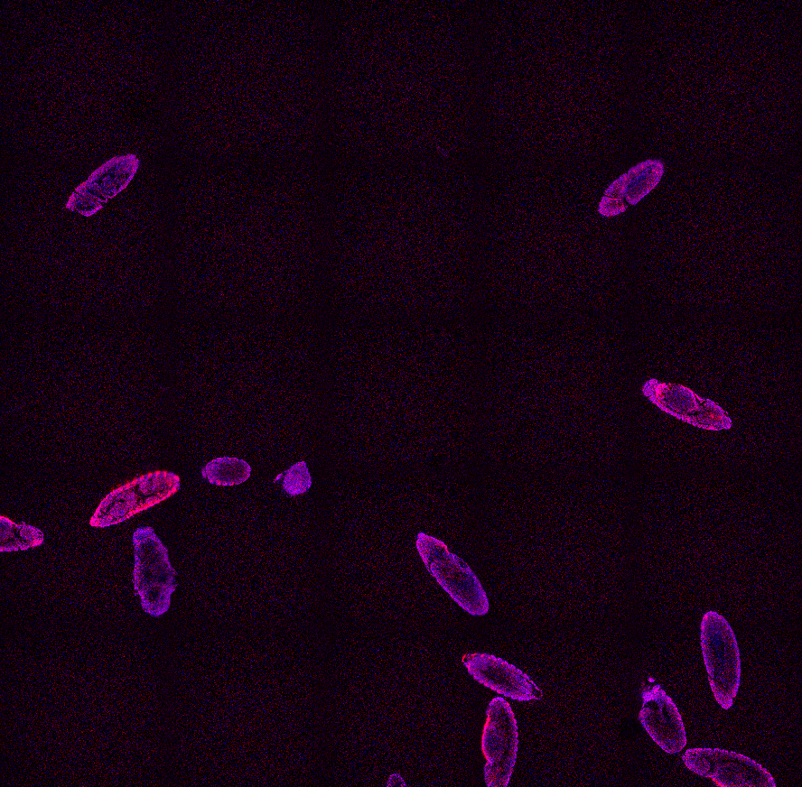
Figure 702. Original image. Courtesy of Richter, Schuetz, Crocker; European Molecular Biology Laboratory, Heidelberg Germany.
We start by defining the following General Analysis:
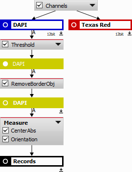
Figure 703. General analysis definition.
DAPI channel is used for segmenting the fruit fly embryos using Segmentation > Threshold > Threshold.
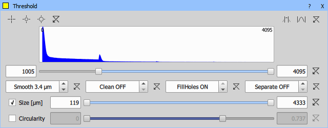
Figure 704. Threshold node.
All embryos touching the image border are then removed using Binary processing > Remove objects > Touching Borders and the center of gravity for each remaining embryo (Measurement > Object position > CenterAbs) and its orientation (Measurement > Object shape > Orientation) is identified. These two variables will be necessary for the scan area positioning.
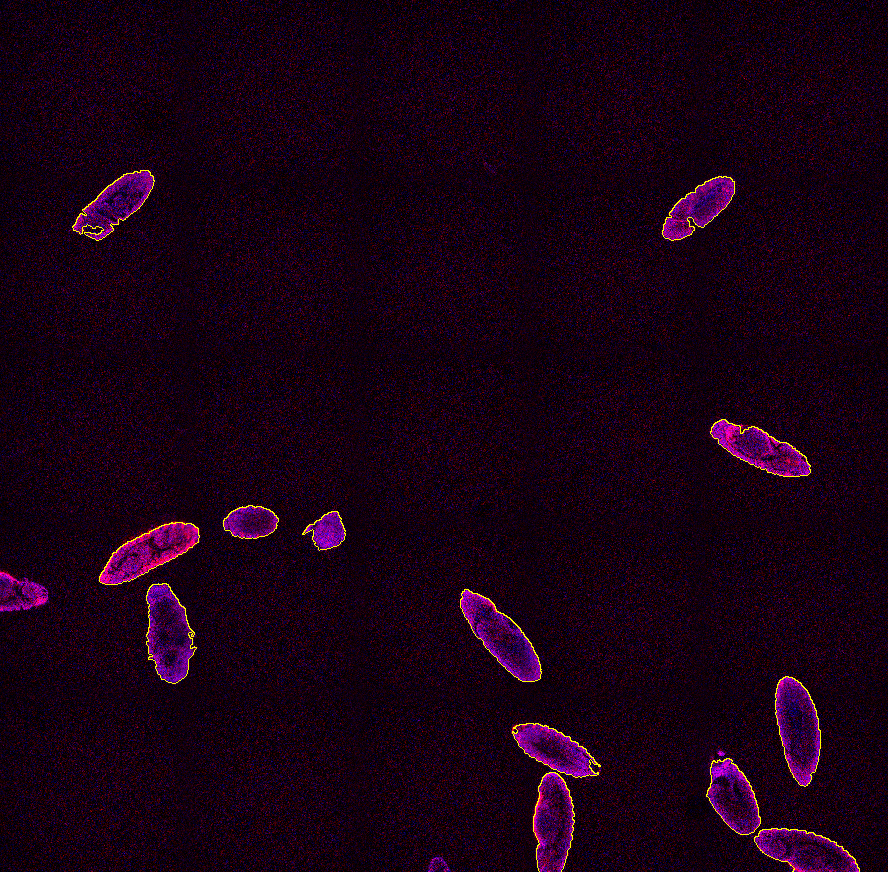
Figure 705. Thresholded embryos.
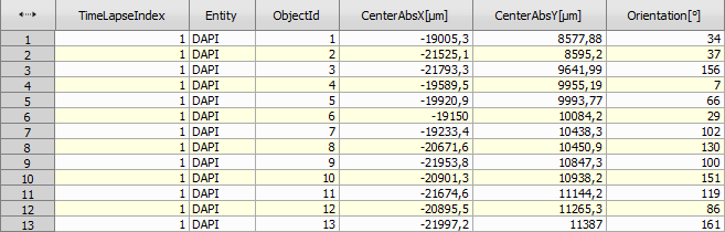
Figure 706. Embryo data generated by the general analysis.
Now we define two optical configurations. One for the low magnification overview (“A1-FULL”) and one for the high magnification scan (“A1-ROTATE”) using the rotated scan area. We make sure Scan Area Settings is unchecked for the latter configuration.
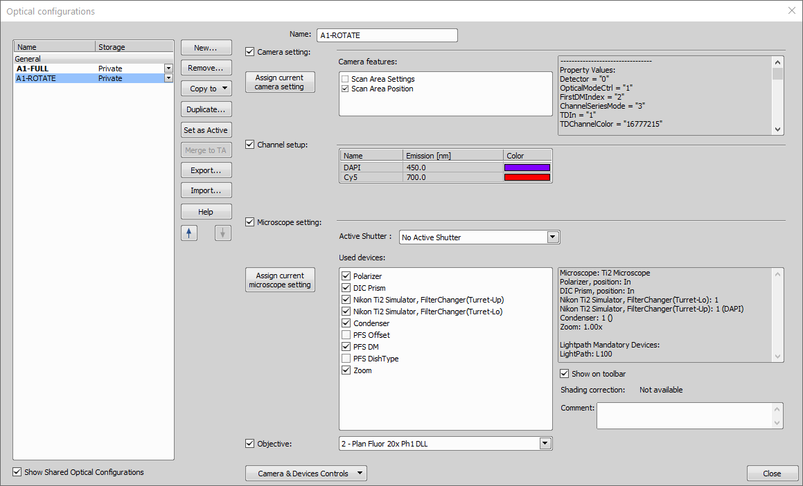
Figure 707. Defined optical configurations.
We can move on by defining a new Job. Low magnification capture is set (Acquisition >  Capture Definition) with the “A1-FULL” optical configuration. Then a new point set (Stage XY Points >
 ![]() New Point Set) is added and filled with the General Analysis 3 data acquired before.
New Point Set) is added and filled with the General Analysis 3 data acquired before.
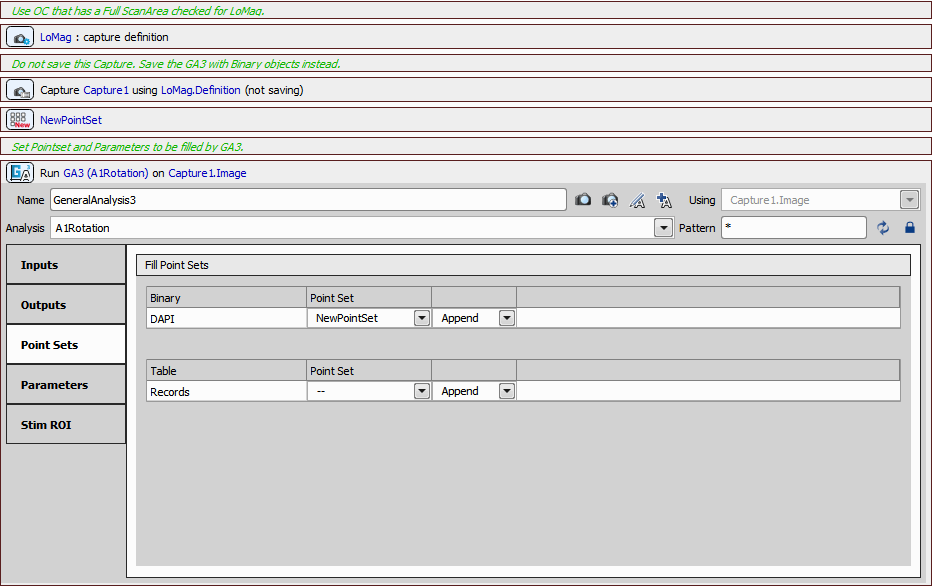
Figure 708. First part of the job definition.
We check all the records available from the selected general analysis (Analysis >  General Analysis).
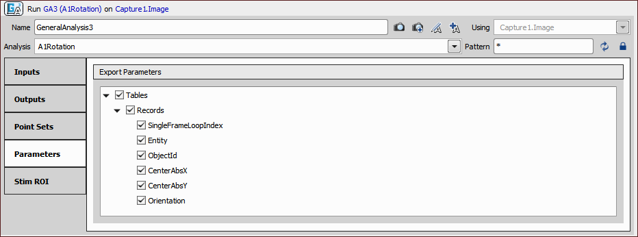
Figure 709. Parameters tab of the General Analysis 3 task.
Then we select the “A1-ROTATE” optical configuration (Optical Configurations >  Select Optical Configuration) and use two macro functions (System >
  Macro) to select the Band Scan scan mode and set the zoom size (8) and shape of the area (2 by 1 rectangle of 1024 x 512).
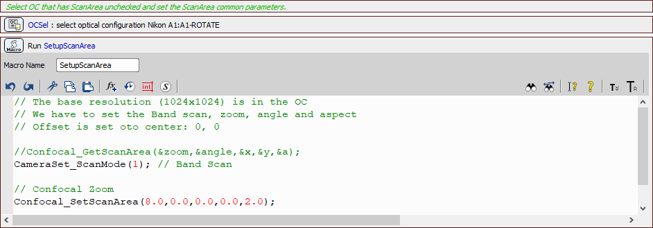
Figure 710. Second part of the job definition.
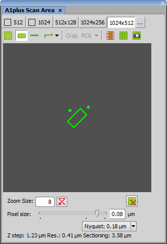
Figure 711. Scan Area settings shown on the microscope pad. These settings will be applied by the job.
Z-Stack for the high-resolution acquisition of the embryo is defined (Z-Stack >  Define Z-Stack) and another macro setting the proper orientation of the scan area is placed inside a point loop (Stage XY Points >
  Loop over Points) together with the Z-Stack loop (Z-Stack >
  Z-Stack Loop) capturing the final images.
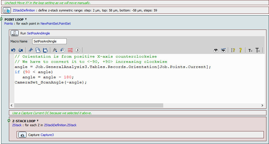
Figure 712. Third part of the job definition.
Now the job is complete and can be executed. Data from the General Analysis 3 are used to properly position the scan area over each embryo and capture the high-resolution Z-Stack.
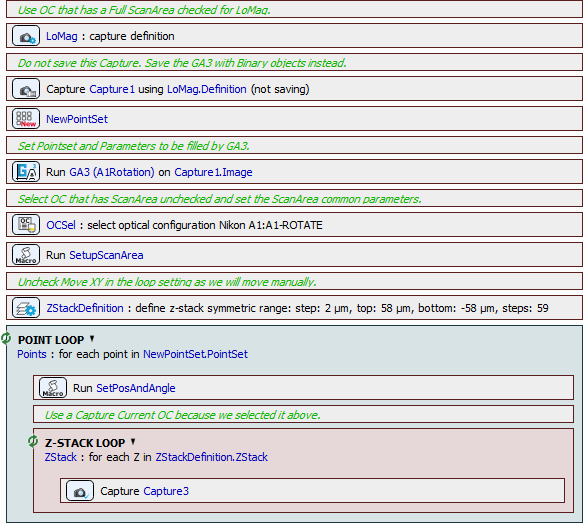
Figure 713. Complete job definition.
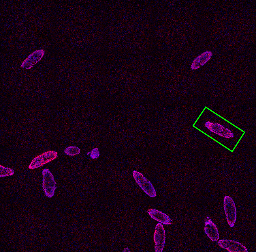
Figure 714. Scan area positioned over one embryo.
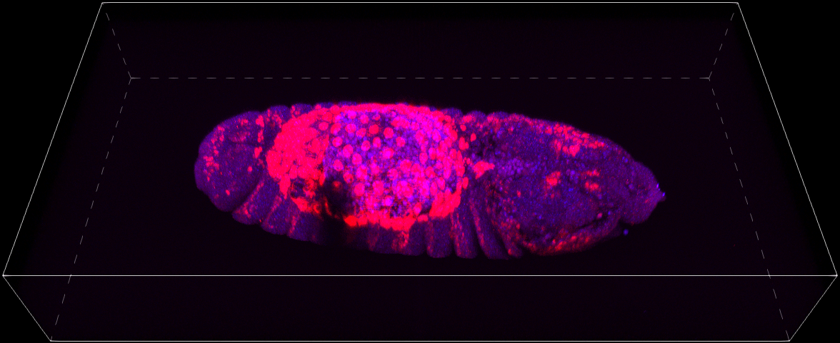
Figure 715. Z-Stack of the same embryo visualized in the Volume Viewer.