In this example, the imaging phase is run over multiple locations at at low rate. When a dividing cell is detected, it is automatically switched to high-speed imaging.
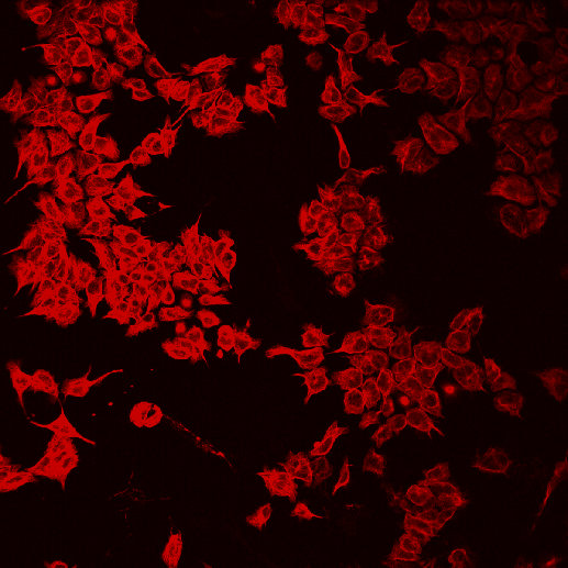
Figure 861. Source image (red is cranked up using LUTs to see the round shape).
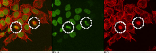
Figure 862. Cells in metaphase.
To detect cells in metaphase using GA3, the following criteria are taken into account:
Cells are small, round and well defined (threshold with restrictions on object size and circularity).
Nuclei are intense and well defined too (spot detection).
Dividing cells have two nuclei.
All these criteria make up the following GA3 recipe:
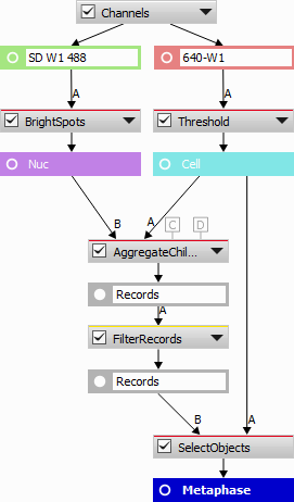
Figure 863. GA3 analysis definition.
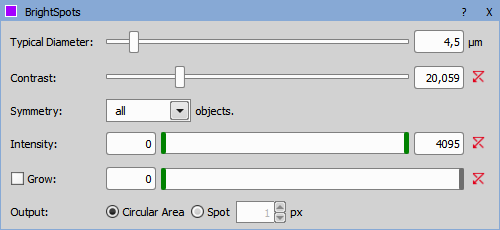
Figure 864. Bright Spots node.
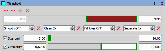
Figure 865. Threshold node.
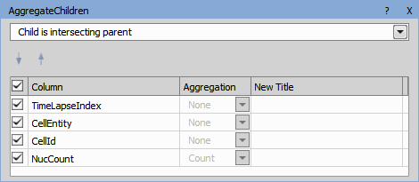
Figure 866. Aggregate Children node.
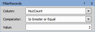
Figure 867. Filter Records node.
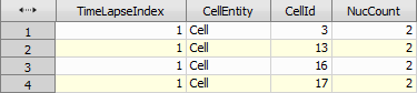
Figure 868. Filtering results.
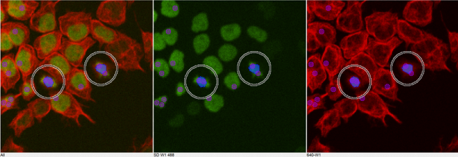
Figure 869. Detected metaphases.
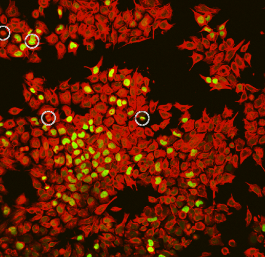
Figure 870. Detection on a different sample.
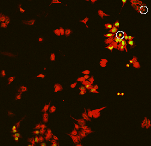
Figure 871. Detection on a different sample.
GA3 in JOBs
Inside JOBs a regular pattern of points on a slide is analysed every 30 minutes using a low magnification objective (“LoMag”). Every image is analysed by the GA3 defined above.The detected cells are then visited and acquired with a high magnification objective (“HiMag”) every minute for half an hour. If no dividing cell is found or after the high magnification acquisition is finished, the experiment goes back to the slow pace.
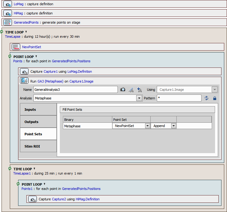
Figure 872. JOBs definition.