
This comprehensive module represents a robust, reliable hardware-software synergy designed to deliver fast, high-quality and efficient microscopic slide scanning in a single, integrated solution.
Key Features
Automated Overview and Metadata
The system automatically captures a full-slide overview, reads sample labels via AI-driven transcription, and ensures all metadata is searchable and organized.
Intelligent Detection and Control
Our AI automatically identifies relevant regions of interest and light settings and still you maintain full manual control to adjust these zones before high-magnification scanning begins.
Precision High-Speed Scanning
By combining Nikon's renowned optics with fast stage technology and precise tiling, you can scan entire tissue sections in a few minutes without compromising on image clarity.
Seamless Multi-Mode Support
The module provides flexibility to switch between brightfield and fluorescence imaging, ensuring the system stays in focus regardless of the sample type or application.
Integrated Quality Control
Transition into a final inspection mode where you can use the smart live tool to compare scanned images side-by-side with the live sample, ensuring total accuracy before finalizing data.
Customizable Results
Organise your data through a flexible cascading tree structure and a convenient gallery view, allowing you to display and modify only the specific information you need for a clean final output.
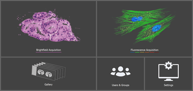
Figure 1528. Home screen.
Home Screen
Predefined brightfield experiment (laser power, exposure, white balance) in which the user simply selects the objective and captures the slides.
Predefined fluorescence experiment for slides capture.

Displays results for all captured slides with a customizable tree structure, allowing users to filter and view only the desired results.

Opens the user management window.

Opens slide scanning settings used for adjusting the holder, image format, shading calibration, filenames, metadata labeling and other global adjustments.